A Beautiful
Canberra Heart Clinic
We Take Care Of Your Healthy Health
Our vision is to bring cardiology services to a whole new level withsophisticated and state of the art technology. With our new and
improved look, we endeavour to provide our patients with the best
quality care and service in a quintessentially beautiful environment.
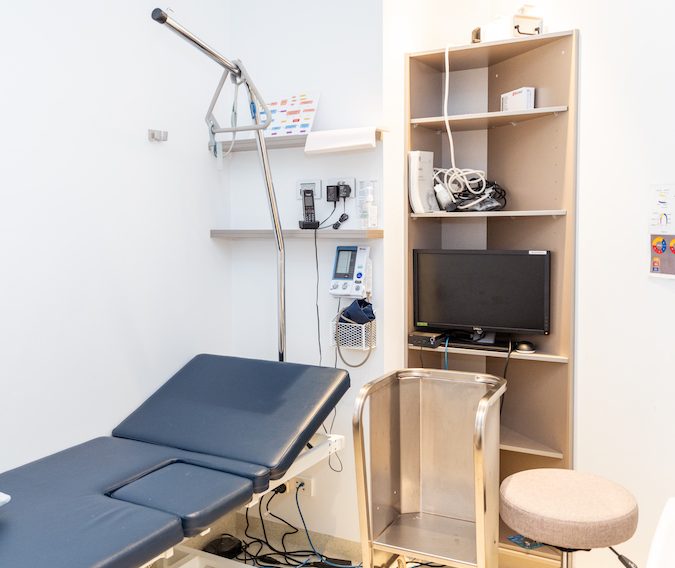
We hope you feel at home in our rooms.Our vision is to bring cardiology services to a whole
new level with sophisticated and state of the
art technology.Make an appointment Make an appointment
Welcome To
Canberra Heart Clinic
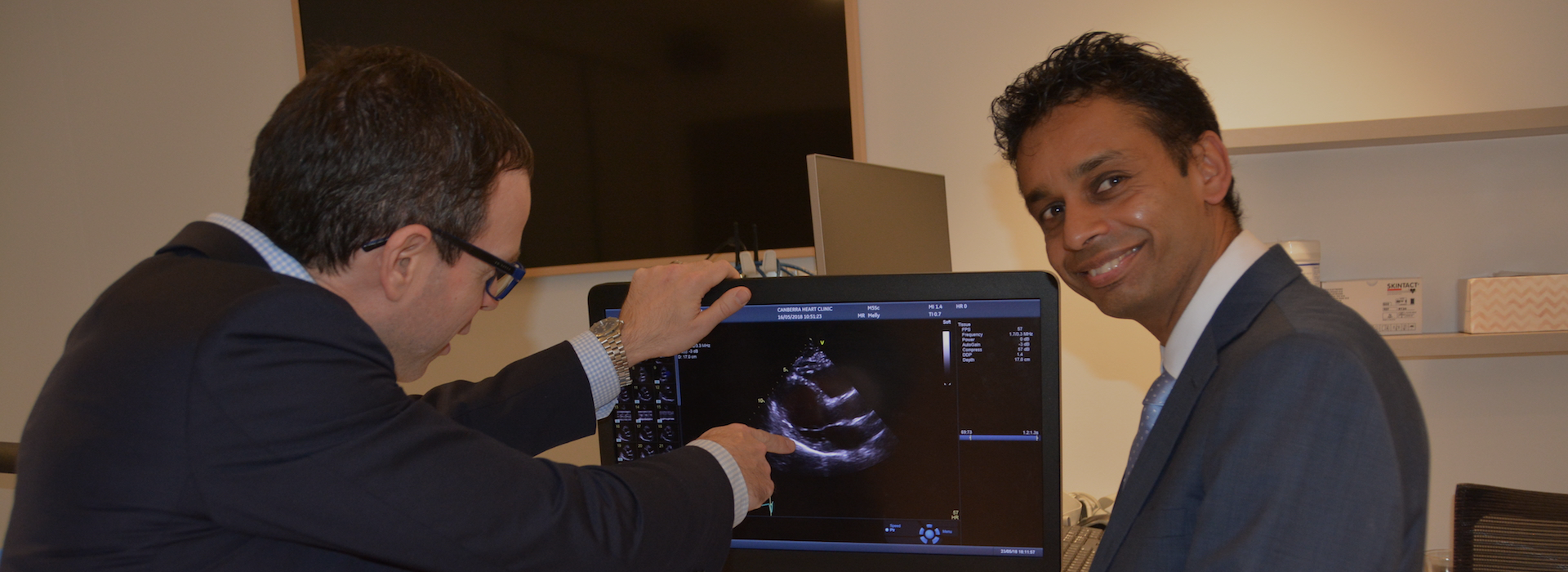
The Canberra Heart Clinic provides expert cardiology consultation, pre-operative assessment, cardiac device follow-up as well as teleconsultation. A comprehensive cardiac investigation service includes transthoracic echocardiography, exercise stress echocardiography, ECG performance and reporting, 24-Hour Holter Monitor to 7-Day Holter Monitor and 24-Hour Ambulatory Blood Pressure Monitoring. Following consultation inpatients procedures if required can be organised. This includes coronary angiography, percutaneous coronary intervention, transoesophageal echocardiography, cardioversion, electrophysiological study and ablation, device implantation, percutaneous structural heart disease therapies and cardiac surgery.
A valid referral is required for all of our services. Healthcare Card discounts are available.
Dr Christopher Allada
Interventional Cardiologist
Director of Canberra Heart Clinic
Canberra heart clinic
Quick Shortcuts
Why Choose Us
What’s Our Resources
Patient Forms
We provide some patient information sheet for you.
- Patient Feedback Form
- Teleconsultation New Patient Worksheet
- Adult Patient Registration Form
- Teleconsultation Review Patient Worksheet
- 7-Day Blood Pressure Diary
- Dizziness Diary
- Paediatric Patient Registration Form

Useful Information
- Daily Progress Tracker
- Canberra Times Articles on Defeat Diabetes
- Weekly Meal Planner with Daily Progress Tracker
- Understanding Carbohydrate Addiction is the Key to Weight Loss
- Training Day - How to Measure Blood Pressure
- Privacy and Access Policy
- Prehydration alone is sufficient to prevent contrast-induced nephropathy after day-only angiography procedures--a randomised controlled trial

To make an appointment with one of our cardiologists:
- How to make an appointment with one of our cardiologists?
- What do I need to do if I cannot make my appointment?
- What do I need to bring to the exercise stress echo?
- Do I need to stop aspirin before my Coronary Angiogram or Coronary Stenting Procedure?
- How long does an Echocardiogram appointment take?
- Do I need a referral to see the cardiologist?


Have Any Question


